
The gait cycle is something you run through 115 to 120 times a minute without giving it a thought. That’s the typical cadence of a young adult, according to a 2017 clinical guide in the Austrian journal Wiener klinische Wochenschrift. Every one of those steps is a precise sequence: the heel lands, the foot flattens, the leg rolls over it, the heel lifts, the toes bend and push, and the foot swings forward to do it all again. Gait labs split that sequence into eight phases and gave each one a name. Knowing them is the foundation for everything else in this section, from why a stiff sole changes your stride to what a flip-flop does to your walk and what your arch is actually for.
The cycle: heel strike to heel strike
The basic unit of walking isn’t the step. It’s the cycle. It starts when one foot touches down and ends when that same foot touches down again. In between, each leg goes through two periods: stance, when the foot is on the ground, and swing, when it’s in the air.
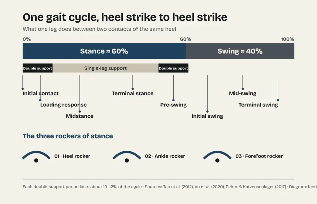
According to a 2020 review of gait phase detection in the journal Sensors, stance takes up about 60 percent of the cycle and swing the remaining 40 percent. Because each leg’s stance periods overlap, there are two moments in every cycle when both feet are on the ground at once. The 2017 guide puts each of those double-support periods at roughly 10 to 12 percent of the cycle. That’s what separates walking from running: when you run, double support disappears and there’s a moment when neither foot is touching the ground.
How fast does all this happen? In 2011, Richard Bohannon and A. Williams Andrews of the University of Connecticut pooled data from 41 studies and 23,111 healthy people walking at their normal pace in a meta-analysis published in Physiotherapy. Average speeds ranged from 1.43 meters per second, about 3.2 miles per hour, for men in their 40s, down to 0.94 meters per second, about 2.1 miles per hour, for women aged 80 to 99. The 2017 guide adds a wrinkle: as people age, they slow down mainly because their steps get shorter, while cadence stays fairly stable.
Eight phases, each with a name
The terminology used in gait labs divides the cycle into eight phases. A 2012 review of gait analysis with wearable sensors by W. Tao and colleagues, also in Sensors, lays them out.
Initial contact is the instant the foot hits the ground, usually heel first. How your joints are positioned at that moment determines how the load gets absorbed. Next comes loading response, the first double-support period, which runs from that contact until the other foot lifts off. The knee bends to absorb the shock, and the ankle lets the front of the foot lower to the floor.
Midstance is the first half of single-leg support. The leg moves forward over a foot that stays planted, while the knee and hip straighten. It ends when body weight is centered over the forefoot. In terminal stance, the heel rises and the leg keeps rolling forward over the front of the foot until the other foot lands. By now the body is out ahead of the forefoot.
Pre-swing is the second double-support period: it begins when the other foot makes contact and ends when this foot’s toes leave the ground. Its job is to set the leg up for swing.
Then come the three swing phases. In initial swing, roughly the first third of the time in the air, the foot lifts and the leg moves forward as the hip and knee bend. In mid-swing, the leg passes the standing leg until the shin is vertical, while the ankle keeps flexing so the foot clears the floor. In terminal swing, the knee finishes straightening and the foot gets ready for a new initial contact.
Three rockers under your foot
During stance, your body doesn’t move over your foot all in one piece. It rolls over it using three different pivot points, which gait researchers call rockers, and which the Tao review places in specific phases.
First is the heel rocker, during loading response: the foot pivots on the heel as the front of the foot lowers toward the ground. Second is the ankle rocker, in midstance: with the foot now flat, it’s the leg that pivots at the ankle, with the shin moving forward over a foot that isn’t going anywhere. Third is the forefoot rocker, in terminal stance: the heel lifts and the whole body pivots over the front of the foot, at the joints at the base of the toes.
The three rockers explain why footwear can change the way you walk: the shape of the heel and the stiffness of the sole come into play right at those pivot points. We dig into the data in our story on heel-to-toe drop.
The windlass that raises your arch
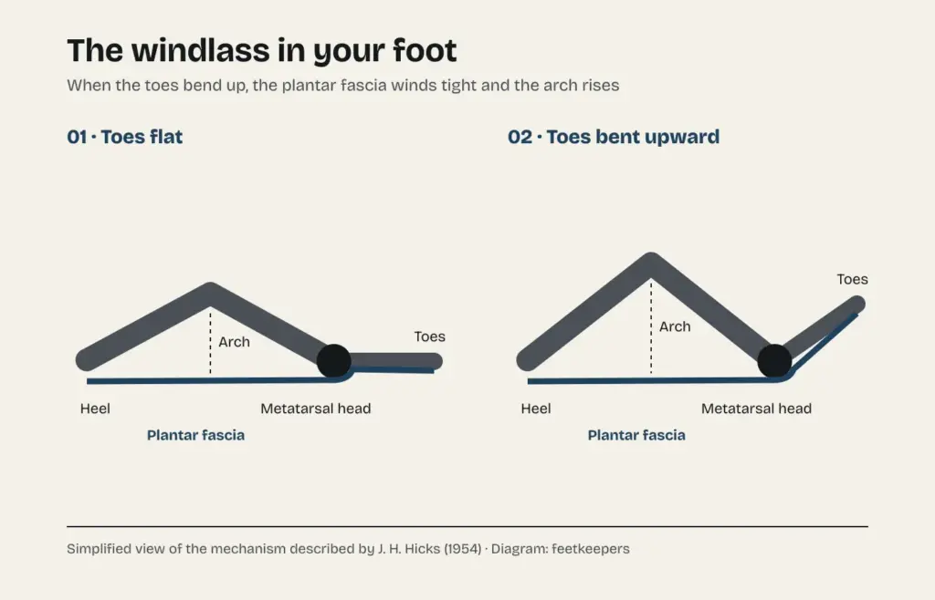
The third rocker hides one of the most elegant mechanisms in the body. In 1954, J. H. Hicks described it in the Journal of Anatomy and named it the windlass, after the drum-and-cable winch used to haul in rope. The key part is the plantar fascia, or plantar aponeurosis, a band of fibrous tissue running along the sole from the heel to the toes.
As a 2020 review in the same journal by F. Sichting, N. B. Holowka, D. E. Lieberman and colleagues summarizes Hicks’s description, the fascia wraps around the heads of the metatarsals, the long bones that end at the base of the toes, like a cable wrapping around a drum, and attaches to the first bones of the toes. When the heel lifts and the toes bend upward, the fascia winds around that drum, tightens and pulls the heel toward the toes. The arch rises and the foot stiffens right when it needs to push off. According to the same review, the mechanism is thought to engage at push-off in walking and running, when the toes bend after the heel comes up.
A 2015 review in the American Journal of Physical Anthropology lists four elements behind a human-style windlass: the calf muscles, the plantar fascia, the inner longitudinal arch and the joints at the base of the toes. The authors call it a significant evolutionary innovation that sets humans apart from other primates, and point out that it’s still unknown when in human evolution it first appeared.
An arch that works like a spring
The arch isn’t just a rigid vault. In 1987, a team led by R. F. Ker and R. McN. Alexander showed in Nature that the arch of the human foot acts like a spring when you run. In the first half of stance, some of the body’s energy gets stored as elastic strain in tendons and ligaments, and in the second half it comes back in the rebound, the way a rubber ball bounces. Calf tendons were already known to play that role. The arch, they showed, does too.
In 2023, an international team led by L. Welte of Kingston, Canada, went further, using high-speed biplanar X-ray, which lets researchers watch the foot’s bones move inside the body. They studied seven people walking and running and compared the real motion of their bones with a model that had no arch recoil. Regardless of how high or low each person’s arch was, that recoil allowed a longer contact time with the ground and better push-off conditions at the ankle for walking upright on a straight leg. The joint responsible, the study found, is one that rarely gets attention: the one between the navicular and the medial cuneiform, two small bones in the middle of the foot. Chimpanzees, the authors note, don’t have that arch mobility during push-off.
The oldest walk on record
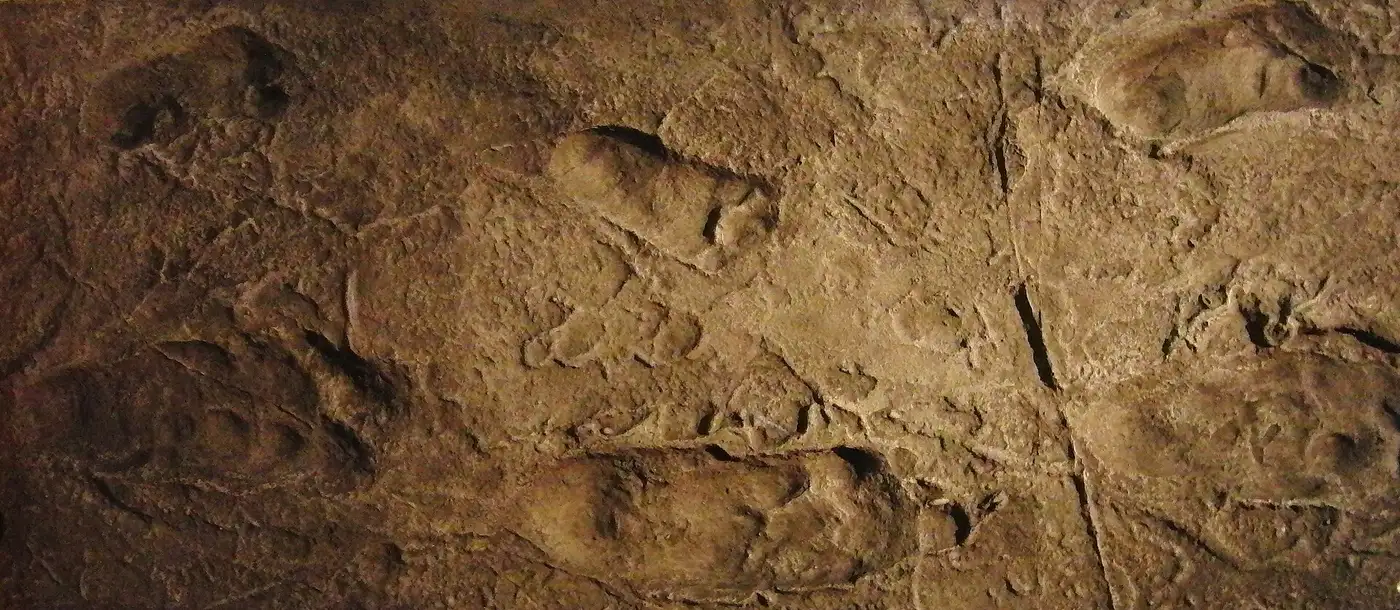
Feet leave a signature on the ground, and some signatures last millions of years. At Laetoli, in northern Tanzania, bipedal footprints discovered in 1978 at what’s called Site G were dated to 3.66 million years ago. They’re usually attributed to Australopithecus afarensis and widely accepted as the oldest unequivocal evidence of obligate two-legged walking in the human lineage.
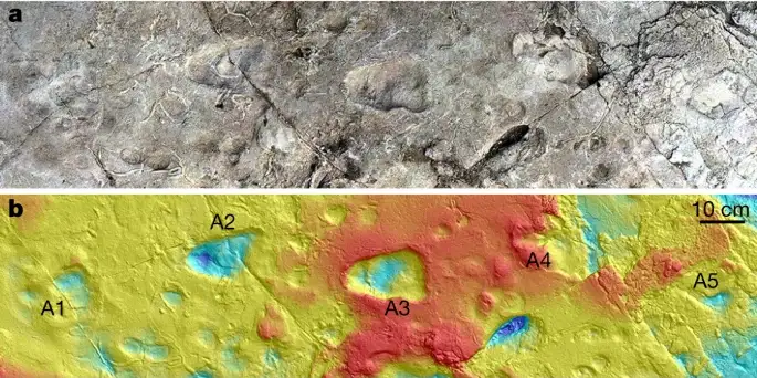
There’s a lesser-known chapter. Two years earlier, in 1976, Peter Jones and Philip Leakey had found five consecutive bipedal footprints nearby at Site A. Mary Leakey tentatively suggested a hominin made them, but their odd shape later led researchers to suspect a bear, and the exact location faded into obscurity. In 2021, a team led by E. J. McNutt and J. M. DeSilva reported in Nature that they had relocated, excavated and 3D-scanned those footprints in 2019. Compared with tracks from black bears, chimpanzees and humans, they looked more like a hominin’s: a small biped that crossed its feet as it walked. And they were different from the Site G prints, suggesting at least two kinds of hominins with different feet and different gaits lived side by side at Laetoli.
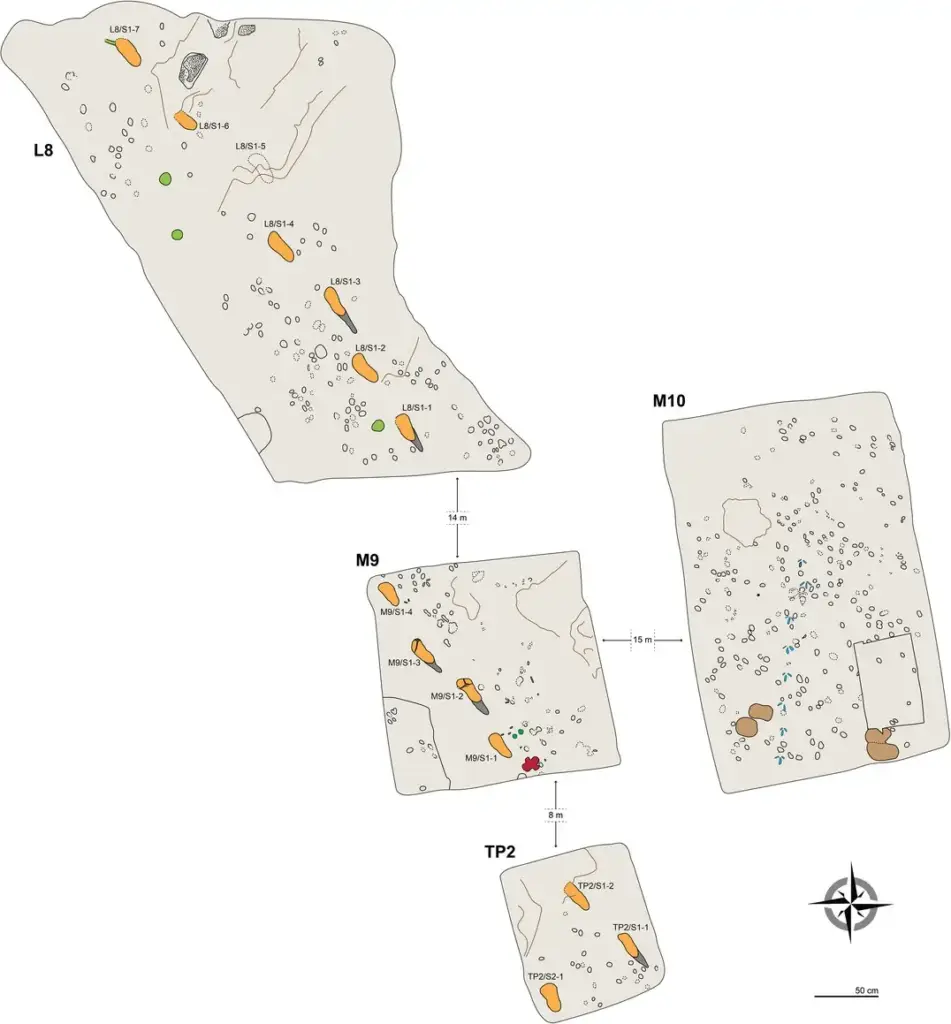
In 2016, an Italian and Tanzanian team published new footprints from a third location, Site S, in eLife. They came from two individuals walking on the same surface and in the same direction as the hominins at Site G. The estimated height of one of them, about 1.65 meters, or roughly 5 feet 5 inches, was far taller than any previous estimate for Australopithecus afarensis.
And in 2023, K. G. Hatala, S. M. Gatesy and P. L. Falkingham showed in Nature Ecology & Evolution that an arched footprint doesn’t by itself prove the foot that made it had an arch. That shape is produced by a particular way of moving the foot that’s characteristic of human walking. The Laetoli tracks show only part of that pattern, with a similar heel strike but a different push-off. The earliest evidence of fully modern human-like walking, by their analysis, comes from early Pleistocene footprints at Ileret, in Kenya, attributed to the genus Homo.
From the page to the camera
Measuring a step precisely took centuries. In 1836, brothers Wilhelm and Eduard Weber published Mechanik der menschlichen Gehwerkzeuge, “the mechanics of the human walking apparatus,” a pioneering study of how we walk. Half a century later, in 1887, Eadweard Muybridge published Animal Locomotion: 781 plates with more than 20,000 photographs, shot in 1884 and 1885 at the University of Pennsylvania, that broke down every phase of human and animal motion into images for the first time.
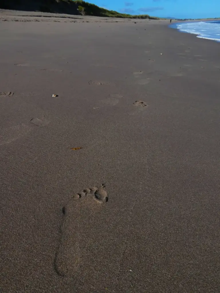
Today gait gets measured with force plates, infrared cameras, sensor insoles, high-speed X-ray and sensors small enough to fit in a shoe, the toolkit the Tao review surveys. The subject hasn’t changed, though: the same eight-phase cycle that leaves its print in the sand on any beach.
What your shoes do with all this
Every part of a shoe comes into play at some point in the cycle. The heel and cushioning act at initial contact and loading response. Sole stiffness affects the forefoot rocker and how far your toes can bend, which is what sets the windlass in motion. How well the shoe holds your instep and heel matters through all of stance, and in swing, the shoe’s weight travels with your leg. That’s why a flip-flop changes how fast your heel comes down, as we explain in our story on flat sandals on cobblestones.
If walking brings on pain that doesn’t go away, that’s no longer a general biomechanics question: see a podiatrist or physician.
Sources
- J. H. Hicks (1954), “The mechanics of the foot. II. The plantar aponeurosis and the arch,” Journal of Anatomy 88(1): 25–30.
- F. Sichting, N. B. Holowka, F. Ebrecht and D. E. Lieberman (2020), “Evolutionary anatomy of the plantar aponeurosis in primates, including humans,” Journal of Anatomy 237(1): 85–104.
- N. L. Griffin, C. E. Miller, D. Schmitt and K. D’Août (2015), “Understanding the evolution of the windlass mechanism of the human foot from comparative anatomy,” American Journal of Physical Anthropology 156(1): 1–10.
- R. F. Ker, M. B. Bennett, S. R. Bibby, R. C. Kester and R. McN. Alexander (1987), “The spring in the arch of the human foot,” Nature 325: 147–149.
- L. Welte, N. B. Holowka, L. A. Kelly, A. Arndt and M. J. Rainbow (2023), “Mobility of the human foot’s medial arch helps enable upright bipedal locomotion,” Frontiers in Bioengineering and Biotechnology 11: 1155439.
- W. Tao, T. Liu, R. Zheng and H. Feng (2012), “Gait analysis using wearable sensors,” Sensors 12(2): 2255–2283.
- H. T. T. Vu, D. Dong, H. L. Cao et al. (2020), “A review of gait phase detection algorithms for lower limb prostheses,” Sensors 20(14): 3972.
- W. Pirker and R. Katzenschlager (2017), “Gait disorders in adults and the elderly: a clinical guide,” Wiener klinische Wochenschrift 129(3–4): 81–95.
- R. W. Bohannon and A. Williams Andrews (2011), “Normal walking speed: a descriptive meta-analysis,” Physiotherapy 97(3): 182–189.
- E. J. McNutt, K. G. Hatala, C. Miller et al. (2021), “Footprint evidence of early hominin locomotor diversity at Laetoli, Tanzania,” Nature 600: 468–471.
- F. T. Masao, E. B. Ichumbaki, M. Cherin et al. (2016), “New footprints from Laetoli (Tanzania) provide evidence for marked body size variation in early hominins,” eLife 5: e19568.
- K. G. Hatala, S. M. Gatesy and P. L. Falkingham (2023), “Arched footprints preserve the motions of fossil hominin feet,” Nature Ecology & Evolution 7: 32–41.
- W. and E. Weber (1836), Mechanik der menschlichen Gehwerkzeuge; E. Muybridge (1887), Animal Locomotion.
Image credits
- Replica of the Laetoli footprints, National Museum of Nature and Science, Tokyo: Momotarou2012. CC BY-SA 3.0, via Wikimedia Commons.
- Gait cycle and windlass diagrams: feetkeepers.
- Laetoli Site A footprints: E. J. McNutt, K. G. Hatala et al. CC BY 4.0, via Wikimedia Commons.
- Map of Laetoli Site S: F. T. Masao et al. CC BY 4.0, via Wikimedia Commons.
- Footprint in the sand, Ireland: Maoileann. CC BY-SA 4.0, via Wikimedia Commons.